Between matter and light - the substance of color. Reseach project in collaboration with Andrew E. Pelling.
Color At Nanoscale
Introduction
The ability to manipulate nature at the atomic and molecular scale is now a reality. Nanotechnology or more aptly put, nanoscience, has become a field that is now entering the public consciousness. Traditionally, this field was defined by the ability to place single atoms and molecules in any desired way but that definition can now be expanded to include the study of diverse topics such as the physical properties governed by the nanoscale size of a material to the motion and mechanical properties of living cells. ìNanoî is the Greek word for dwarf and has come to identify the scale on which all these phenomena take place: 0.000000001 meters. We cannot see objects at this scale with our eyes but special microscopes have been developed to feel surfaces at the nanoscale and provide a tactile interface between the scientist and the atom. Scientists have discovered that the properties of a material at this invisible scale control its properties at the macroscale. For example, the way atoms are arranged in a metal will affect the structural properties of that metal when it is used as a support structure in a bridge. The strength of that metal arises from its atomic makeup. Nanoscience holds the potential to change technologies we use today and also has applications in manufacturing, medicine, fashion and art.
Quantum dots
Quantum dots are one such material in which their macroscale properties are governed by their nanoscale makeup. Quantum dots semi-conductor particles that are highly fluorescent and whose spectral properties depend on their small size. The colour they appear is determined by quantum mechanics, hence the name ìQuantum Dotî. Simply put, quantum mechanics is the study of matter and radiation (light) at the atomic scale. It is no surprise then, that the properties of nanoscale materials are related to their quantum mechanical properties. In a quantum dot, electrons are confined into a very small space. When this happens the energies of the electrons become discreet and they are only allowed to take on certain values. For instance the energy of an electron might have a value of E = n, where E is its energy and n = 0, 1, 2, 3, and so on. In this case it is impossible for the electron to have the energy E = 1.5 since n can only be an integer. This is analogous to a walking up a staircase; it becomes impossible to try and stand in between the individual steps. In quantum dots, n is partly defined by the size of the quantum dot and in turn this is related to its colour.
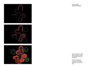
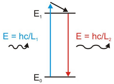
In order for a material to be fluorescent it must be able to absorb light of a specific wavelength and then emit light at a different wavelength. Light is made up of particles called ìphotonsî which carry energy that is related to their wavelength by the formula E = hc/L, where L is the wavelength, c is the speed of light and h is a universal constant. The wavelength of light that can be absorbed is proportional to the size of the discreet energy steps of the material. When a quantum dot absorbs a photon, it moves into an excited energy state and some of the energy the light carries will be dissipated, as heat or vibrations (in general), and the rest will be released as a new photon with a slightly lower energy as the system relaxes back to its ground state (figure 1). This process is known as fluorescence when the photons the quantum dot emits reaches our eyes we see a brightly coloured material.
Figure 1. Fluorescence is the process by which a material will absorb a photon and release a new photon with a lower energy and longer wavelength. An incoming photon is absorbed from the ground state of the material (E0) and causes the material to go into an excited energy state (E1) (up arrow). The energy between E0 and E1 is discreet and any excess energy the incoming photon was carrying will be dissipated as heat or vibrations (diagonal arrow). As the system relaxes back to its ground state (down arrow) it will release a new photon that will have less energy and a longer wavelength than the original photon.
Seeing the invisible
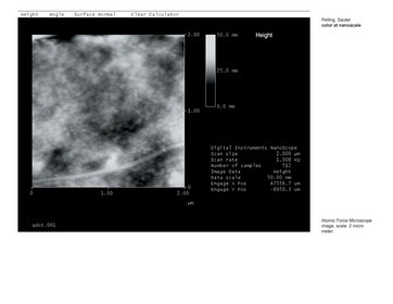
When billions and billions of quantum dots are placed on a surface we can easily see the photons they emit with a traditional optical microscope. However, in order to visualize quantum dots at the nanoscale, a special type of microscope is needed. The Atomic Force Microscope (AFM) does not use light to ìseeî things but rather relies on tactile sensing. A cantilever, with a small tip at the end can be mounted on a piezoelectric crystal that can move in the X, Y, and Z directions. If the tip is scanned over a surface, the probe moves up and down over its topography and the displacement caused by features on the surface can be measured to create an image. The movement of the tip is very similar to the way a needle in a record player moves over bumps in a record. By manufacturing very small tips, it is possible to image atoms and molecules on a surface. This microscope represents a paradigm shift in microscopy, from seeing with our eyes to ìseeing by feelingî.
The interface between the visible and the invisible becomes the realm from which colour and light originate. The AFM provides a unique mechanism by which we can explore the relationship of light and matter at the nanoscale.
The Concept
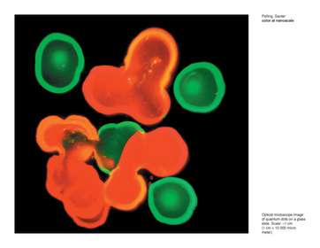
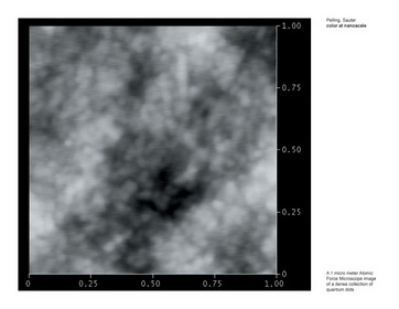
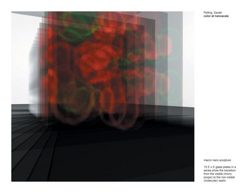
To explore the interface between light, scale and colour we propose a series of images captured with the optical microscope and the AFM. Quantum dots can be dried onto a glass slide and AFM images can be collected. AFM imagery reveals the nanoscale topography of the quantum dots as we zoom from 10 micrometers to 1 micrometer (1 meter = 1 million micrometers). At a 1 micrometer scan size the individual quantum dots begin to become visible. At a larger scale (1 centimeter = 10000 micrometers) the quantum dots on the glass slide can be excited with white light and images can be captured with an optical microscope and the bright colours of the quantum dots become apparent. Without the white light exciting the quantum dots, they are a dull colour and are barley visible on the glass slide. In the following pages we present typical images of the quantum dots captured with these methods and propose a public installation/sculpture that explores the origin of light at the nanoscale.
download research document (pdf, 1 MB)
| 1
| 2
| 3
| 4
| 5
| 6